TMJ disorders are a common orofacial pain condition affecting millions of individuals worldwide. Accurate assessment and diagnosis are crucial for effective treatment planning. While clinical examination remains essential, imaging techniques provide invaluable insights into the complex anatomy and pathology of the TMJ. This paper discusses various imaging modalities used in the evaluation of TMJ disorders, including panoramic radiography, cone-beam computed tomography (CBCT), magnetic resonance imaging (MRI), and arthrography. Additionally, the the benefits and limitations of each technique and emphasizes the role of interdisciplinary collaboration for optimal patient care.
Temporomandibular joint (TMJ) disorders encompass a range of conditions affecting the complex temporomandibular system, causing pain, dysfunction, and compromised quality of life. Evaluating TMJ disorders poses a significant diagnostic challenge due to the intricate anatomy and dynamic nature of the joint. Clinical examination alone may not provide a comprehensive understanding of the underlying etiology and structural abnormalities. Therefore, imaging techniques play a vital role in assessing the TMJ, aiding in accurate diagnosis and subsequent treatment planning.
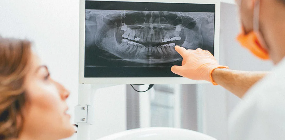
- Panoramic Radiography: Panoramic radiography is a commonly employed imaging modality for initial TMJ evaluation due to its accessibility, low radiation dose, and ease of use. It provides an overview of the condyle, mandibular fossa, and surrounding structures. However, panoramic radiographs have limitations in depicting detailed TMJ anatomy and assessing soft tissue abnormalities. It may not be adequate for complex cases requiring comprehensive evaluation.
- Cone-Beam Computed Tomography (CBCT): CBCT has gained popularity in TMJ imaging due to its superior spatial resolution and ability to provide multiplanar reconstructions. CBCT enables precise visualization of osseous structures, condylar position, articular surfaces, and TMJ morphology. It is particularly useful in assessing bony abnormalities, such as condylar resorption, osteoarthritic changes, and internal derangements. However, CBCT involves a higher radiation dose compared to panoramic radiography, necessitating judicious use.
- Magnetic Resonance Imaging (MRI): MRI is considered the gold standard imaging modality for TMJ evaluation. It provides excellent soft tissue contrast, allowing visualization of the articular disc, joint effusion, synovial membrane, and surrounding muscles. MRI is essential in diagnosing internal derangements, disc displacement, and assessing the presence of joint inflammation or degenerative changes. Additionally, it aids in differentiating TMJ disorders from other orofacial pain conditions. However, MRI is relatively expensive and not widely accessible in some regions.
- Arthrography: Arthrography involves injecting contrast material into the TMJ to enhance visualization during imaging. This technique provides dynamic information about disc position and function, making it valuable in diagnosing internal derangements. Arthrography can be performed using different imaging modalities, such as fluoroscopy or MRI. It helps determine appropriate treatment options, including arthrocentesis, arthroscopy, or surgical interventions. However, arthrography is an invasive procedure and carries potential risks and limitations.
In conclusion, imaging techniques are indispensable tools in the evaluation and diagnosis of TMJ disorders. While clinical examination forms the foundation of assessment, imaging modalities, including panoramic radiography, CBCT, MRI, and arthrography, offer crucial information to accurately identify the underlying etiology and guide treatment decisions. Each imaging technique has its strengths and limitations, necessitating a judicious selection based
TMJ Treatment by Dr. Nojan Bakhtiari
Dr. Nojan Bakhtiari is a highly experienced cosmetic dentist and one of the leading providers of Botox for TMJ and headaches in New York City, NY. With years of experience and a commitment to providing the highest quality care, Dr. Bakhtiari is an expert in the use of Botox for the treatment of TMJ disorder and headaches.
Dr. Bakhtiari has a deep understanding of the underlying causes of TMJ disorder and headaches, and has extensive experience in using Botox to treat these conditions. He uses a combination of the latest techniques and technologies, including Botox injections, to provide his patients with the most effective treatment possible.
If you are looking for a safe and effective way to treat your TMJ disorder or headaches, Dr. Bakhtiari and his team are here to help. With their expertise and commitment to providing the highest quality care, you can trust that you will receive the best possible treatment for your condition.
To learn more about Botox & TMJ treatment by Dr. Nojan Bakhtiari & schedule your consultation today!