The temporomandibular joint (TMJ) is the joint that connects the jawbone to the skull and is responsible for enabling movement of the jaw. TMJ disorders are conditions that affect the normal functioning of this joint and can lead to pain, discomfort, and difficulties in jaw movement. Some of the most common symptoms of TMJ disorders include jaw pain, popping or clicking of the jaw, difficulty in opening and closing the mouth, and headaches.
Diagnosis of TMJ disorders is crucial to ensure that the right treatment can be prescribed. With an accurate diagnosis, patients can receive the necessary care to alleviate their symptoms and improve their quality of life. In this article, we will look at the various methods used to diagnose TMJ disorders and the role of Dr. Nojan Bakhtiari in the diagnosis and treatment of these conditions.
Diagnostic Methods for TMJ Disorders
The first step in diagnosing TMJ disorders is a thorough evaluation of the patient’s symptoms, medical history, and oral habits. This will help the doctor determine if the symptoms are related to the TMJ and to identify any underlying causes of the condition.
Physical Examination
The physical examination is a crucial step in the diagnosis of TMJ disorders. During the examination, the doctor will assess the patient’s jaw movement, listen for any clicking or popping sounds, and check for any tenderness or pain in the jaw area. The doctor may also perform a range of other tests, including range-of-motion tests, muscle strength tests, and tests that assess the patient’s bite and jaw alignment.
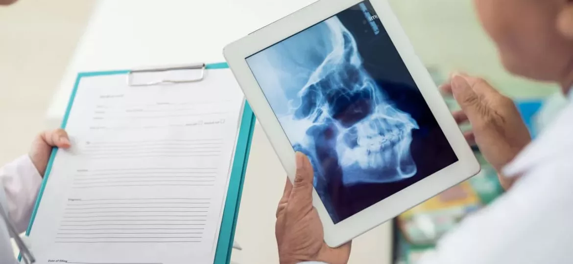
Imaging Tests
Imaging tests are often used to get a better understanding of the anatomy of the TMJ and to identify any structural abnormalities that may be contributing to the patient’s symptoms. Some of the most commonly used imaging tests include X-rays, CT scans, and MRI scans.
X-rays are useful for evaluating the bone structure of the jaw and TMJ, and can show any signs of degeneration or damage to the joint. CT scans are more detailed and can show more precise images of the bones and soft tissues in the area. MRI scans are the most advanced imaging tests and are used to evaluate the soft tissues and muscles of the jaw, providing detailed images of the entire joint.
Arthroscopy
Arthroscopy is a minimally invasive surgical procedure that allows the doctor to visualize the inside of the joint and assess any damage or abnormalities. During the procedure, a small camera is inserted into the joint through a small incision, and the doctor can observe the joint and surrounding structures on a monitor. This procedure is especially useful in cases where the patient has already undergone imaging tests and the doctor needs a more precise evaluation of the joint.
Diagnostic Models
Diagnostic models are casts or models of the jaw that can be used to evaluate the patient’s bite and jaw alignment. These models are usually made by taking an impression of the patient’s upper and lower teeth and using the impression to create a cast or model of the jaw. The doctor can then use the model to assess the patient’s bite and jaw alignment and determine if any adjustments need to be made to improve function.
The Role of Dr. Nojan Bakhtiari in TMJ Disorders Diagnosis
Dr. Nojan Bakhtiari is a highly skilled and experienced dental specialist who is dedicated to helping patients with TMJ disorders. He has extensive training in the diagnosis and treatment of these conditions and uses a range of diagnostic tools and techniques to help identify the underlying cause of a patient’s symptoms.